Persyst Video Library
- Persyst 15 Overview (26:26)
- Persyst Mobile Overview (14:23)
- Persyst Trending Overview (6:19)
- Persyst Artifact Reduction (3:28)
- Seizure Review (12:52)
- Spike Review (9:54)
- FFT Spectrogram – Part 1 (9:48) and Part 2 (7:16)
- Z-Score comparison to patient’s Baseline (19:57)
- Suppression Ratio Trend Panel and creating Notifications (7:05)
- Propagation Maps (7:56)
- Modified Channel Sets (11:39)
- Creating a Time Trend (6:17)
- Creating and Exporting Spike Groups (9:15)
- Creating and Saving Custom Trends (8:06)
- Topographic Brain Mapping (19:31)
- Persyst 14 Seizure Detection Settings (8:37)
- EEG Electrical Source Imaging (ESI) for Epilepsy Evaluation Overview (15:51)
Persyst Quick Reference Document Library
- Creating Custom Montages
- Creating Custom Electrode Maps
- Creating Frequency and Voltage Topographic Maps
- Persyst Database Configuration
- Event Density Tool
- Calculating Spike Wave Index
- Exporting Spike Detections
- Creating Custom Filters
- High Frequency Display
- Full Neoprism Guide
- Shared Settings
- Clip Archive Quick Guide
- Persyst 14 Research Overview
- Persyst 13 Comment Filters
- Persyst 14 Back Averaging
- Persyst 15 Quick Reference Guide for a Reduced Set of Electrodes
Webinar recordings hosted by Persyst
- Persyst 14; Quantitative EEG for cEEG with speaker Michael Guess.
- Using Persyst 14 in the EMU. Recorded September 23, 2020 with speaker Dr. Mark Scheuer, M.D.
- Using Persyst 14 in the ICU. Recorded November 4, 2020 with speaker Dr. Mark Scheuer, M.D.
- An expert panel discussion on EEG Electrical Source Imaging (ESI). Recorded March 17, 2021 with speakers Dr. Stephan Schuele, M.D., M.P.H., Dr. Stephan Bickel, M.D. Ph.D., & Dr. Leonardo Bonilha, M.D., Ph.D.
Persyst FAQ
See something missing? Ask us to add it at support@persyst.com.
- Verify that study is processed
- Click on Track Acquisition and compare time shown in trends to time at end of study
- Manually check for trend results files in study location
- Change sensitivity to High
- This may indicate whether anything was marked at lower perception values
- Verify any issues with the recording workflow
- Verify that study was recorded using 10-20 channel labels
- Verify that study does not have a large amount of open channel EEG or calibration in first 5 minutes of recording
- Manually identify something that should have been marked and contact support@persyst.com.
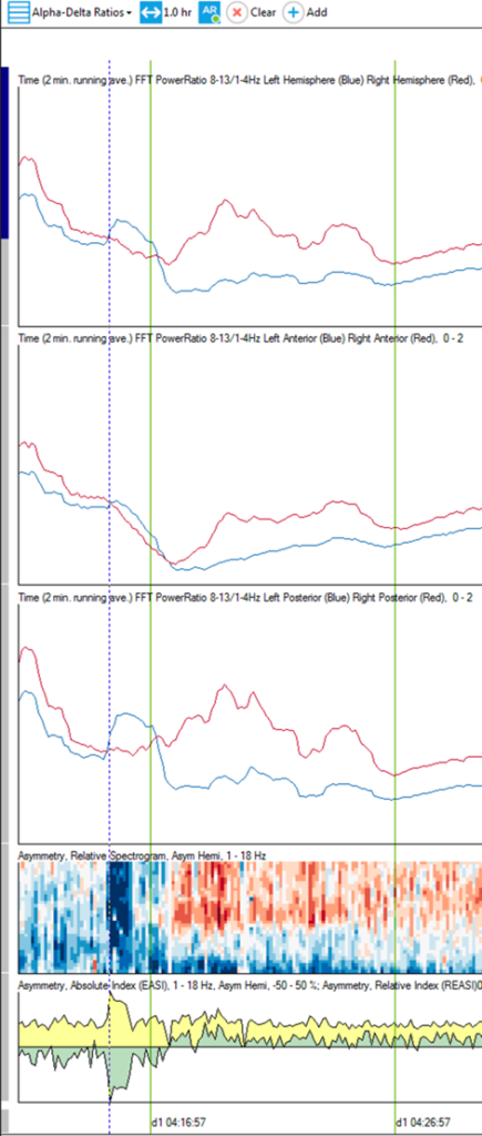
The Alpha/Delta ratio panel would be helpful for examining regional slowing. In this screenshot from the primary study that we looked at yesterday you can see that following the seizure (which starts at the dotted line cursor) the slowing on the left (blue line in the ADR trends) is much more pronounced compared to the right (red line).

Yes. Persyst Artifact Reduction isolates and selectively reduces muscle artifact. Even though the motor activity might be a visual cue for the seizure onset, the Persyst seizure detector is focused on the underlying EEG patterns, not on the muscle activity.
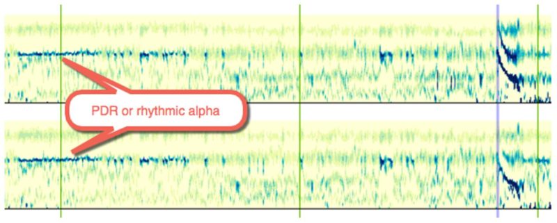
The Rhythmicity spectrogram clearly shows PDR.

PHI (Protected Health Information)
Persyst software is a medical device. Just as a heart rate monitor has access to PHI, Persyst Software has access to PHI. Also, like a heart rate monitor, the data collected / analyzed resides entirely within your control at all times and like a heart rate monitor manufacturer, Persyst Development Corporation does not have access to PHI.
Check registration and license type
- Launch Persyst standalone to verify the program opens
- Check for correct background color and .mmx file to indicate whether Deploy Settings was run.
- If Persyst opens, navigate to Preferences | General Preferences | About and check the type of license (EMU/AMB licenses do not support trending).
- If the software is properly registered, verify that OEM software read the registration correctly.
- Natus: Check for any “Persyst License Error” annotation in Natus annotation list.
- Nihon Kohden: Verify that launching MMFrame does not display any errors and that there are no black lines diagonally across the MMFrame Window
Check for correct configuration (situational by OEM)
- Natus: verify that analyzer is present in Tools | Options | Analysis tab and pointed at the correct .mmx
- Nihon Kohden: verify that the correct status, file path, and executable option are selected
- Nicolet: Verify that the Nicolet dongle has the Persyst option licensed and that the Nicolet Integration was run during install
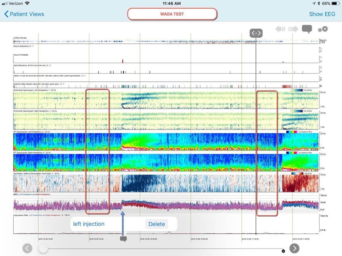
(a.k.a. Amytal Study or intracarotid sodium amobarbital procedure (ISAP))
A combination of trends on the Comprehensive Trend Panel are best for observing the slowing and return to baseline. On the screenshot below the left injection is marked and also a segment before the injection and one just before the right injection where the EEG looks to have returned to the baseline prior to the injection.

- Go to Start | All Programs | Persyst (or Insight) | RegCenter.
- Select “Request Activation License” (or “Save to File” on older versions).
- Fill in all of the information. In the field marked “Registration Number” insert your Persyst Software Registration Number (which should be on your CD box or in download email).
- Press “OK” and you will be prompted to save the file. Save this file in a location where you can find it, or to a removable media that you can take to a computer with Internet access.
- Email the file to support@persyst.com
Persyst will email back an unlock key. Simply move this key file to the system you are unlocking and double click on the file. You will be asked if you want to add the information to the registry. Select “Yes” and you should receive a confirmation that the information was added to the registry. Please be aware that you will need administrative rights to install and unlock the software.

