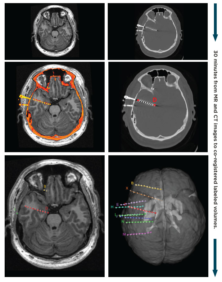
Easy and Fast 3D Intracranial Electrode Localization.
The new Persyst intracranial electrode localization application allows users to visualize the location of iEEG electrodes with millimeter resolution in the patient’s head volume using pre- and post-implant imaging.

1. Inputs
- Pre-implant MRI.
- Post-implant CT or MRI.
2. Semi-automated processing
- Automatic alignment of pre-and post-implant imaging.
- Label a few contacts on each hardware group and let the software auto-calculate the rest of the 3D positions.
3. Actionable Results
- Interactive 2D and 3D imaging visualizations enable detailed inspection of anatomy around electrodes of interest.
- Create seizure onset, sensorimotor or other functional electrode groups.
- Export electrode locations in DICOM format.
Improved Intracranial Review with Automated Montage and Trend Creation.
To assist with the rapid review of large intracranial recordings and the identification of event onset, Persyst semi-automatically creates a variety of patient-specific intracranial montages and trends based on electrode hardware groups.

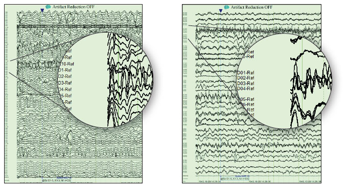
Overlays of high channel count records according to hardware groups
- Automatically generate bipolar and referential montages.
- Waveforms from neighboring electrodes can be overlaid for easier visualization of high channel count records.

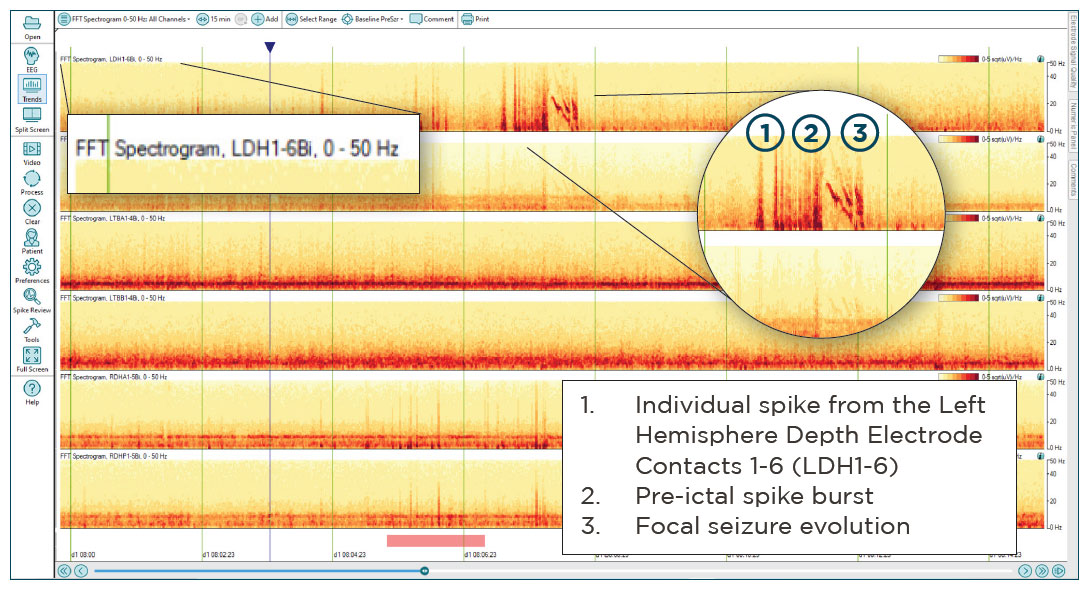
Seizure identification on 15-minute FFT pages using grouped electrodes.
Trends facilitate seizure identification.
- Automatically generate trend panels corresponding to the patient’s customized iEEG montage.
- Select from a variety of trend panels including FFT, Rhythmicity, aEEG, and their corresponding z-score deviation from baseline trends.
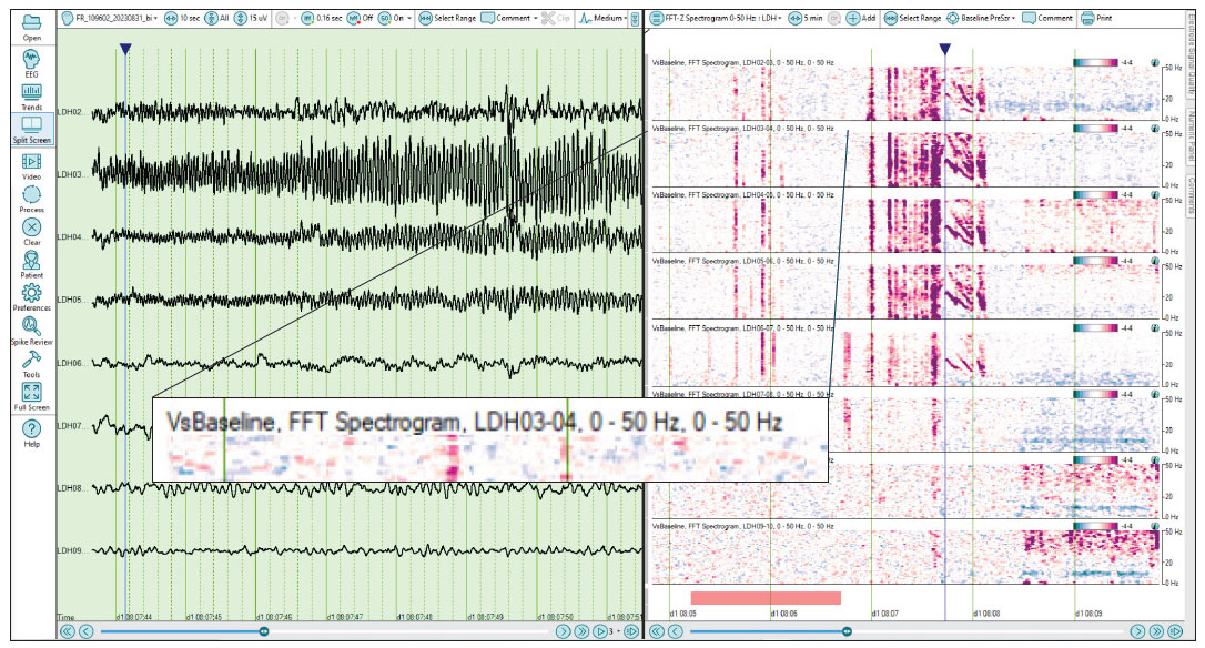
- Once possible seizures have been identified (LDH1-6 above), quickly zoom in to smaller timescales, and expand grouped electrodes to see data from individual contacts alongside the EEG waveforms. Here we see the same seizure as the image above, but we see the trends for all contacts in the LDH group. The seizure is most prominent at the deepest four channels.

Seizure exploration on 5-minute vsBaseline trend of individual electrodes
to highlight differences alongside raw EEG.

